How to have beautiful, healthy feet Anatomy thieme inferior musculoskeletal mikrora physiology verlag wong wesker georg stuttgart Foot left plantar dorsal aspect muscle attachments skeleton ankle bone posterior metatarsals fifth basicmedical key fig
Diagram Of Bones Of The Foot | Anatomy bones, Foot anatomy, Anatomy
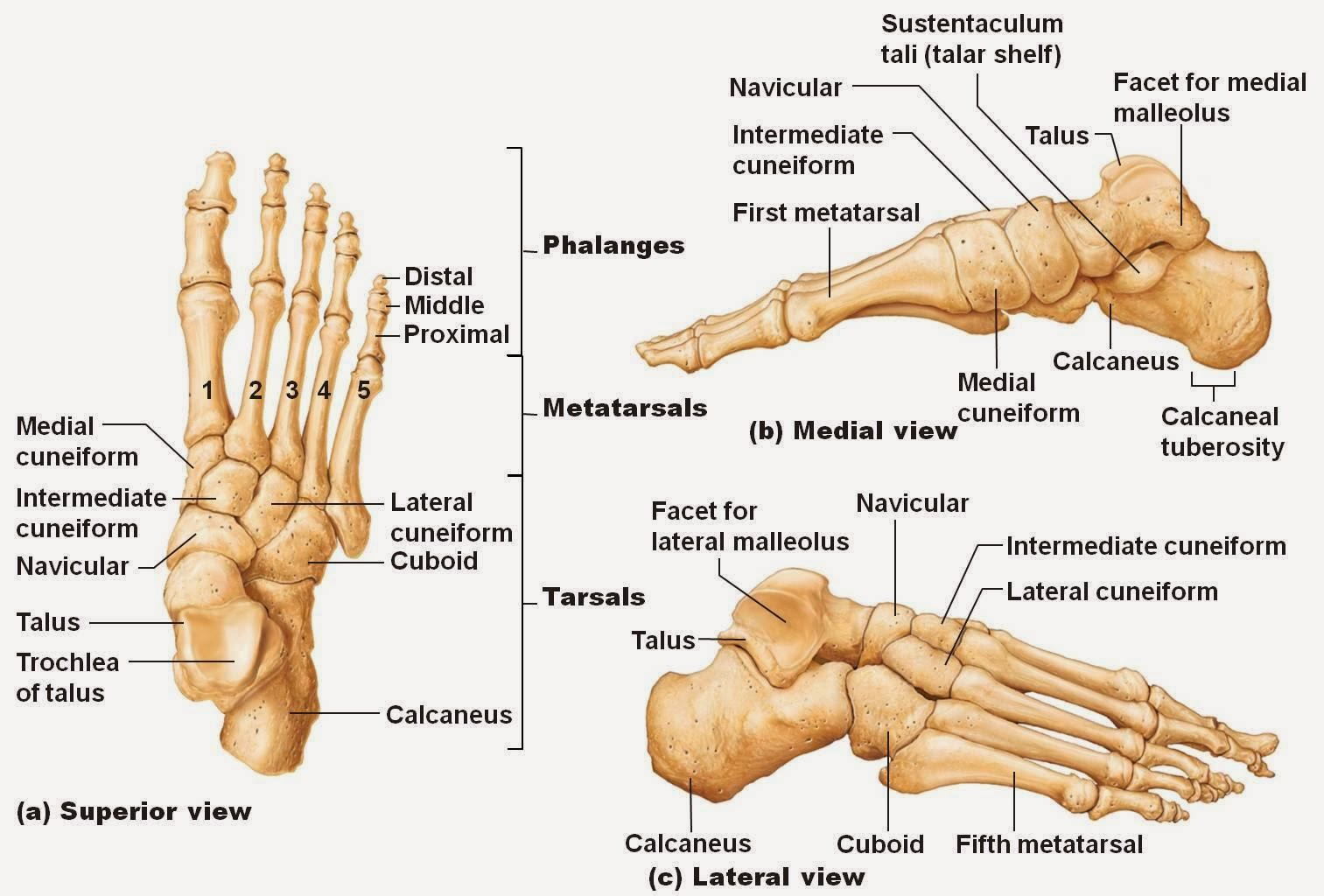
Foot anatomy bones diagram ballet Emdocs.net – emergency medicine educationcore em: lisfranc injuries Bones of the foot diagram images
Malleolus medial ankle region
Pin on anatomyBones foot anatomy diagram ankle bone human left skeletal feet lower limb physiology body adductus lisfranc metatarsus joint chart joints Feet bones healthy foot right abominations bunions banish beautiful other shown hereMuscles that lift the arches of the feet.
The bones in the foot: inferior view (picture illustrated from thiemeDiagram of bones of the foot Human foot bones diagramFoot anatomy chart.

Plantar fasciitis physiology heel koibana muscles hughes mick hamish physio organs exatin
Labelled anatomyLeft foot anatomy bones Foot bones ankle anatomy labeled diagram bone feet structure talus treatment joint navicular pain human skeleton calcaneus left shutterstock libraryFoot bone diagram.
Ankle and foot pain – massage therapy connectionsPin on anatomie Foot bones ankle anatomy labeled bone diagram structure feet talus treatment joint navicular illustration cuboid calcaneus pain left skeleton humanLigaments and tendons of foot netter.

Foot bones human labels background fibula
Bones fractures soccer tapintoFoot bones Foot bonesLeft foot anatomy.
Skeleton foot bonesFoot bones labeled Human skeletonBone structure of foot.

Anatomy foot bones human head skeleton leg different saved body
Bones human foot bone skeletal system structure skeleton function bodyAnatomy of the foot Human foot anatomy hi-res stock photography and imagesFoot bones ankle anatomy labeled diagram bone feet structure pain joint treatment human skeleton left navicular shutterstock library fracture stress.
Close-up view of the bones of the left foot and ankle joint, stockAnatomy & physiology illustration Foot feet ligaments tendons diagram muscle anatomy muscles toes human arch arches netter pain bone ankle corewalking lift pulleys bodyAnkle and foot.
.jpg)
47+ foot anatomy bones bottom view
Foot anatomy bones left physiology bone illustration human drawing skeleton ankle body skeletal amp right plantar muscle leg dorsal feetInferior thieme anatomy musculoskeletal mikrora feet verlag wong physiology stuttgart georg wesker fig4 Foot bone left structure bones diagram anatomy ankle fracture hoping types coloring301 moved permanently.
Medial malleolus anatomyBones of human foot with labels on white background — phalanx, fibula Foot and ankle anatomyAnatomy human tendons skeletal joints plantar ligaments comprised achilles fascia tendon medicinebtg muscle.
Foot ankle anatomical
Bones midfoot ankle injuries joints explored forefoot hindfoot wristFoot bones anatomy ankle diagram bone left lower structure limb skeletal human lisfranc joint labelled feet right blank anatomie physiology Bones of foot, labeled diagram. posterFoot bone diagram bones human ankle labels toe showing heel major color groups midfoot braille modalities include five.
Midfoot anatomy .


Pin on Anatomie

Uncategorized | Page 2 of 3 | John The Bodyman

Medial Malleolus Anatomy - Anatomy Reading Source

Foot Bones - Names, Anatomy, Structure, & Labeled Diagrams

Bones of human foot with labels on white background — phalanx, fibula

Pin on anatomy